Retinal detachment repair
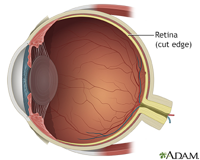
Normal anatomy
|
|
The retina is the internal layer of the eye. It receives and transmits images that have passed through and been focused by the lens and cornea.
|
Indications
|
|
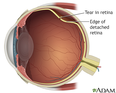
Retinal detachments are associated with a tear or hole in the retina. The internal fluids of the eye may leak through the hole or tear, causing the retina to separate from the tissues underneath. Risk factors for retinal detachment include age-related eye changes, eye injury or surgery, nearsightedness, and diabetes. Symptoms of retinal detachment include bright flashes, floaters, or loss of part of the visual field. Emergency retinal detachment surgery is necessary to prevent vision loss.
|
Procedure, part 1
|
|
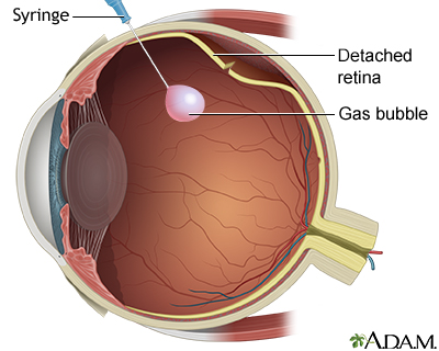
The two most common methods of repairing a retinal tear are pneumatic retinopexy and placement of a scleral buckle. Pneumatic retinopexy is often used for smaller detachments near the front of the eye. In pneumatic retinopexy, a gas bubble is injected into the back of the eye.
|
Procedure, part 2
|
|
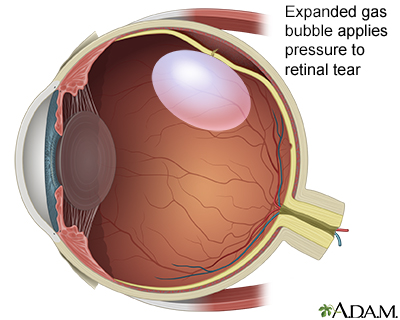
The gas bubble expands and pushes the retina back in place against the wall of the eye.
|
Procedure, part 3
|
|
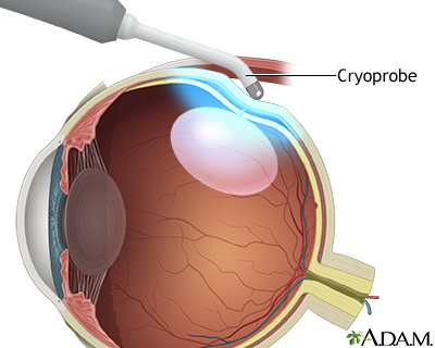
A small scar is created that will cause the two layers to seal together as the scar develops. There are two main ways to do this: cryopexy and photocoagulation. During cryopexy, a cryoprobe is used to freeze the tissue in area of the retina that has the tear. The freezing process causes a scar to form..
|
Procedure, part 4
|
|
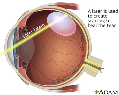
Photocoagulation uses a laser beam passed through the front of the eye and focused on the area around the tear. The tiny laser burns the tissue, which causes scarring. The choice of which to use, cryopexy or photocoagulation, is often determined by location of the tear (front or back part of the retina) and the amount of space between the detached retina and the underlying tissues.
|
Procedure, part 5
|
|
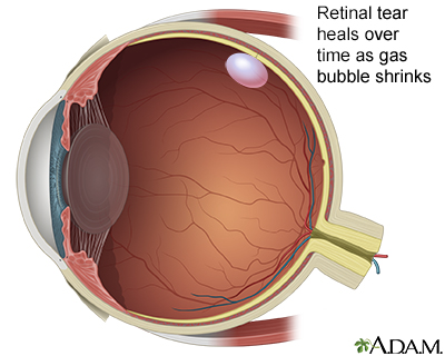
Over time, the retinal tear heals as the gas bubble shrinks and eventually goes away. The gas bubble will usually last about a week, and the person may have to stay in the proper position for the entire time to keep the bubble in place.
|
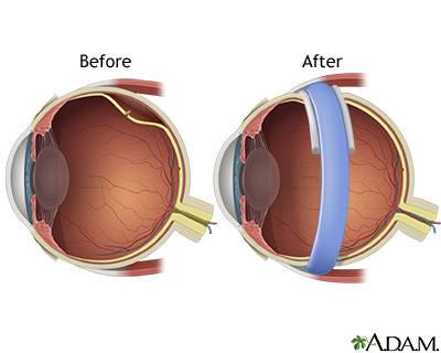
Procedure, part 6
|
|
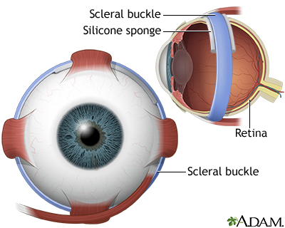
The second method to bring the layers together is placement of a scleral buckle. A scleral buckle is often used for retinal tears and detachments that require more advanced surgery. After sealing breaks and tears in the retina using either a cryoprobe or photocoagulation, a silicone sponge is sewn to the outside of the eye. This pushes the wall of the eye inward to meet the detached retina. This compresses the globe of the eye, which may elongate slightly. Sometimes a silicone band is wrapped around the eye, and over the sponge (like a belt), so that it pushes the sponge in a little more. The silicone sponge and band (if used) usually can be left in place permanently, unless they cause problems later, such as infection.
|
Aftercare
|
|
Scleral buckling for detachment may require a short time in the hospital but people often can go home the same day. It is important to keep the head elevated at all times. The person should not bend over or strain when lifting or with bowel movements. Vigorous exercise should be avoided for 3 to 4 weeks.
|

Review Date:7/24/2025
Reviewed By:Franklin W. Lusby, MD, Ophthalmologist, Lusby Vision Institute, La Jolla, CA. Also reviewed by David C. Dugdale, MD, Medical Director, Brenda Conaway, Editorial Director, and the A.D.A.M. Editorial team.
The information provided herein should not be used during any medical emergency
or for the diagnosis or treatment of any medical condition. A licensed medical professional
should be consulted for diagnosis and treatment of any and all medical conditions. Call 911
for all medical emergencies. Links to other sites are provided for information only -- they
do not constitute endorsements of those other sites. © 1997-A.D.A.M., Inc. Any duplication or distribution of the information contained herein is strictly prohibited.
The Agency for Health Care Administration (Agency) and this website do not claim the information on, or referred to by, this site is error free. This site may include links to websites of other government agencies or private groups. Our Agency and this website do not control such sites and are not responsible for their content. Reference to or links to any other group, product, service, or information does not mean our Agency or this website approves of that group, product, service, or information.
Additionally, while health information provided through this website may be a valuable resource for the public, it is not designed to offer medical advice. Talk with your doctor about medical care questions you may have.